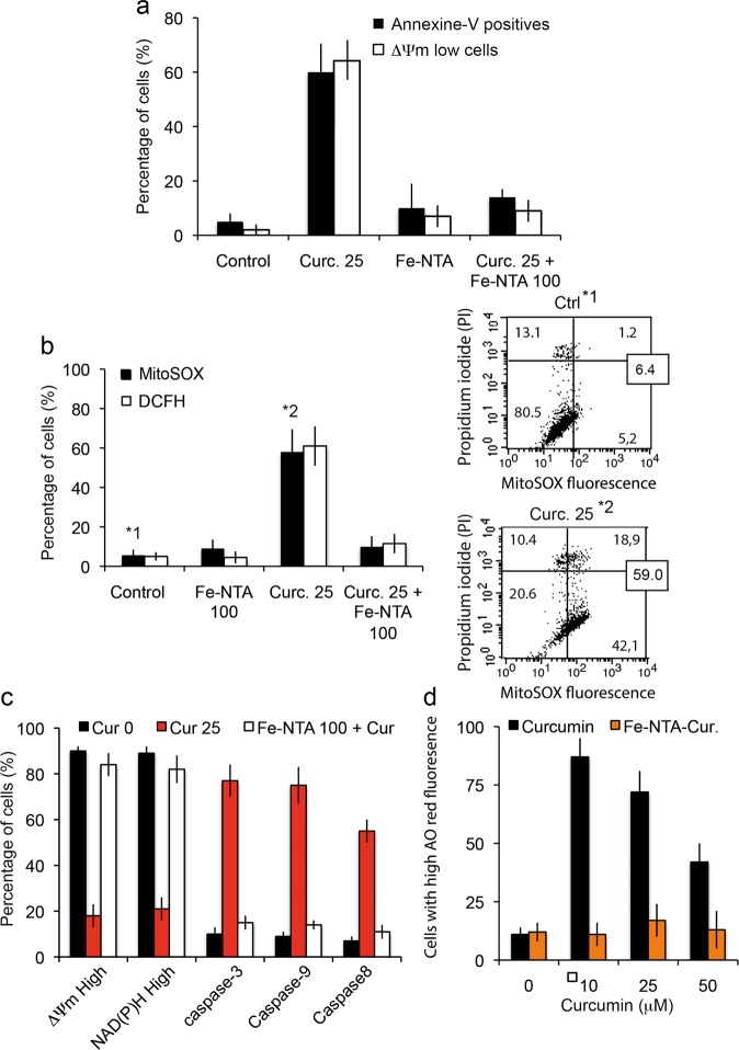
Fig. 6. Cell death induced by curcumin and Fe-NTA-curcumin complex.
a Flow cytometry analysis of early and late events during cell death (respectively, drop in mitochondrial membrane potential (ΔΨm) and PS exposure) when cells are treated for 24 h with curcumin (20 μM) or Fe-NTA (100 μM) alone or with both, which behave intracellularly as chelators. The results are issued from 7 independent experiments. b Flow cytometry analysis of the events following the ΔΨm drop (in a): generation of superoxide anions (MitoSOX staining) and hydroperoxide production (DCFH-DA staining. Each histogram is from seven independent experiments and ±SD is given. Control and 20 µM curcumin (24 h) conditions are illustrated respectively in 1* and 2* by biparametric plots together with the viability measurement (PI staining). The percentage of cells in each panel is given as a percentage (%) of the whole population. c Broad metabolic analysis showing that treatment with both products restores control conditions, with respectively a large population of high ΔΨm and NAD(P)H together with low activation of the so-called “initiation caspase” (caspase-8) and “executioner caspases” (caspase-3, -7 and caspase-9). Cells were treated with 20 µM curcumin or 100 µM Fe-NTA or both. d Cells were treated for 24 h with curcumin or Fe-NTA, or both. Then we analyzed red acridine orange fluorescence. As curcumin alone induces low pH vesicle staining which can be related to autophagy induction (more lysosomes and appearance of autophagic vesicles,) the Fe-NTA-curcumin complex abolishes that increase and returns to baseline level.

