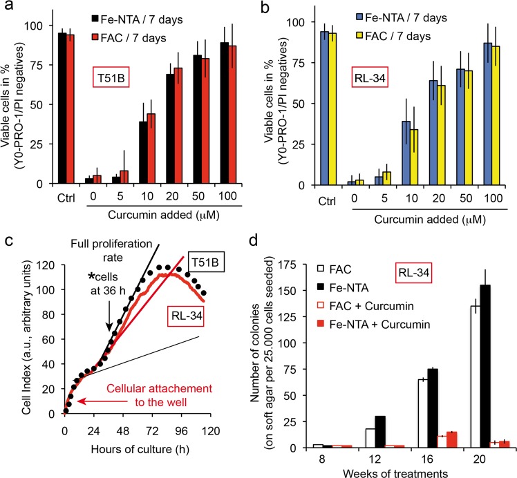
Fig. 7. Curcumin chelate with free iron alleviates the tumor-promoting activity of Fe-NTA or FAC.
a T51B cells are protected against free iron toxicity by curcumin in a concentration-dependent manner. Epithelial T51B cells were treated for 7 days with either 100 µM Fe-NTA or 100 µM FAC ( + 10 µM hydroxyquinoline) and various curcumin concentrations and then viability-tested by cytometry taking into account the dead cells (apoptotic, A and necrotic, N cells) to find the cellular viability [100 − (A + N) = viable cells in percentage]. b Same as in a, but with RL-34 cells. c The cells used in the c experiment were taken at 36 h in their full proliferation phase after plating. The situation was tested by impedancemetry using the ACEA Xcelligence system. Arrows (black) indicate the situation of the cells at 36 h and the arrow (in red) early in the curve indicates the initial cell binding to the wells, which in our case took almost 24 h before full proliferation. The curve is drawn taking into account the mean value for 12 different wells. d T51B cells plated at 2.105 cells in 60 mm dishes were treated with MNNG (0.5 mg/mL for 24 h) after plating and cultured with either 20 μM curcumin or Fe-NTA 200 μM (or FAC 250 μM with 8-hydroxyquinoline), or both. The culture medium was renewed every 2–3 days with fresh media. T51B cells were trypsinized before they became confluent (day 4–6). The culture continued for up to 20 weeks and colony formation was tested every two weeks, special care being taken when formation became greater at 16, 18, and 20 weeks of culture). The number of soft agar experiments conducted was n = 7 and ± SD are indicated. MNNG was added at the start to participate in the initiation of tumorigenesis.

