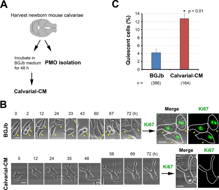
Figure 1.
Calvarial conditioned medium (Calvarial-CM) confers cellular quiescence to C4-2B4 PCa cells. (A) Calvariae prepared from 2–5 day-old newborn mice were cultured in BGJb medium containing 0.1% BSA for 48 h to generate Calvarial-CM. Calvariae were also used to isolate primary mouse osteoblasts (PMOs) (see details in Materials and Methods). (B) Live-cell imaging analysis of C4-2B4 PCa cells incubated in media containing control BGJb media or Calvarial-CM. Single cells were monitored on a Nikon BioStation and images were acquired every 20 min for 72 h. (Left) Phase contrast brightfield images. Arrowheads follow one control cell through three cell divisions. Round cells are undergoing mitosis. Note that one daughter cell left the field of view after T = 33 h. (Right) Immunofluorescence images. Immediately following time-lapse, cells were fixed and immunostained for the proliferation marker Ki67 and re-imaged on the BioStation. Phase contrast images are merged with immunofluorescence images for Ki67. Cell outlines are traced for ease of view. All bars, 20 µm. (C) Quantification of % quiescent C4-2B4 cells that did not divide over 72 h relative to total cells examined (mean ± s.e.m.). n, number of cells monitored. N, number of independent experiments for control BGJb (N = 5), Calvarial-CM (N = 4). P values were by t test.

