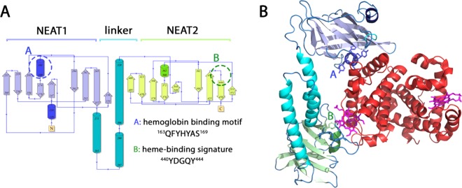
Figure 1.
IsdB secondary and tertiary structures. (A) Topology of IsdB (PDB code 5vmm). The structure consists of 18 β-strands and 6 α-helices, forming two NEAT domains connected by a flexible linker. (B) 3D cartoon structure of wt IsdB in complex with methemoglobin (MetHb). Hb binding motif and heme binding signature are represented in stick residues. A and B refer, respectively, to the hemoglobin binding motif and to the heme-binding signature.

