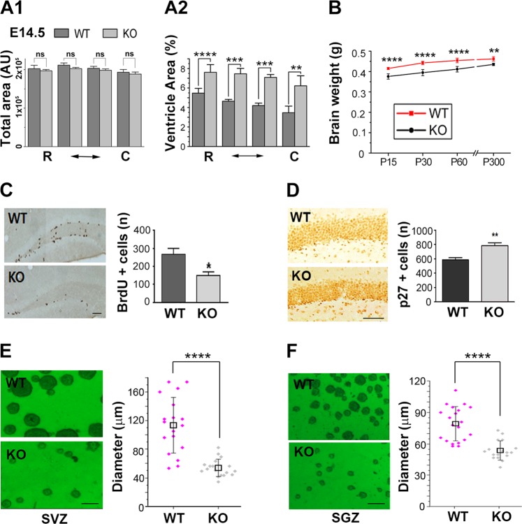
Fig. 6. PARP-1 KO mice exhibited reduced brain weight and decreased proliferation of adult NSCs.
a Total brain area (A1) and ventricle area were measured from Nissl-stained brain sections of PARP-1 littermates at ED 14.5 and the percent cross-sectional ventricle area over total brain area (A2) was quantified (n = 5 for each group). b Brain weights of littermates were measured at postnatal day 15 (WT, n = 12; KO, n = 12), day 30 (WT, n = 9; KO, n = 13), 60 (WT, n = 7; KO, n = 7) and day 300 (WT, n = 5; KO, n = 4). c Immunolocalization of proliferating adult progenitor cells in the hippocampal dentate gyrus from WT (n = 5) and PARP-1 KO (n = 4) mice (scale bar = 50 μm) and quantitative analysis of BrdU-positive cells. d Immunohistochemical localization of p27-positive cells in the dentate gyrus from WT and PARP-1 KO mice and quantitative analysis of p27-positive cells (n = 4 for each group). e, f Neurosphere cultures derived from the adult SVZ (e) and SGZ (f) of WT and PARP-1 KO mice (scale bar = 100 μm) and the sizes of neurospheres were compared (e; WT, n = 38; KO, n = 33, f; WT, n = 40, KO, n = 35; pooled from four independent cultures). Means ± SD are shown (*p < 0.05, **p < 0.01, ****p < 0.0001, two-tailed unpaired t-test).

