Figure 3.
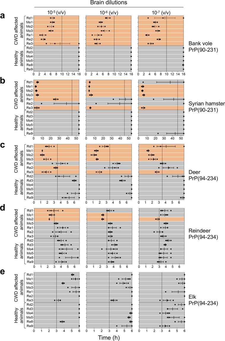
RT-QuIC results of CWD affected animals with detectable PrPSc in the brain. RT-QuIC analysis of serial brain homogenate dilutions (from 10−5 to 10−7) from CWD affected animals and controls with (a) bank vole, (b) Syrian hamster, (c) deer, (d) reindeer and (e) elk recombinant truncated PrP. Each sample was analyzed in triplicate and black dots indicate the time taken for each replicate to reach the fluorescence threshold (lag phase). The vertical line indicates the time threshold set up for each PrP substrate. Rd: red deer, Mo: moose; Re: reindeer. Mean value and standard error of the mean (S.E.M) are shown.

