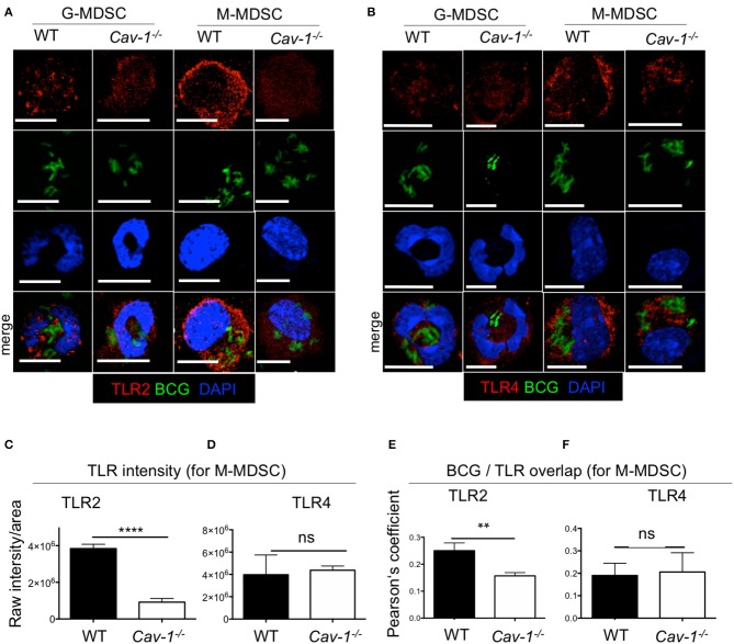
Figure 2.
Cav-1 deficient MDSCs show reduced intracellular TLR2 but not TLR4 in BCG-infected vesicles. MDSCs were stimulated with BCG-GFP for 16 h at 1 MOI. Cytospins were stained for TLR2 (A) or TLR4 (B) and analyzed by confocal microscopy. G-MDSCs and M-MDSCs were identified on the basis of polymorphonuclear or mononuclear shape by DAPI staining. All scale bars 11μm. (C,D) Quantified data of raw intensity/area for TLR2 or TLR4. of M-MDSCs from WT and Cav1−/− mice. (E,F) Pearson's correlation coefficients for colocalization of BCG with TLR2 overlap and BCG with TLR4 overlap. Data represent for (C) n = 30 cells, for (D) n = 30 cells, and for (E,F) n = 10 cells. ****P < 0.0001; **P < 0.01; ns, not significant unpaired, two-tailed, student's t-test.

