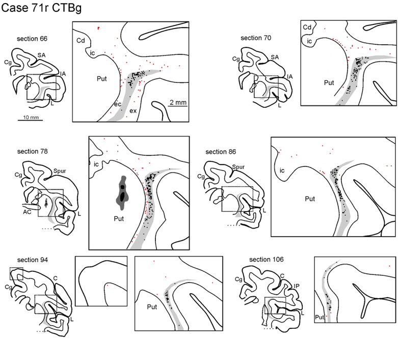
Figure 3:
Drawings of coronal sections showing the distribution of the labeled neurons observed in the WM and the claustrum in Case 71r CTBg. The sections are shown in an anterior (section 66) to posterior (section 106) order. For each AP level, the section outline shows from where the enlargement was taken (box). In the enlargements, red dots correspond to retrogradely labeled WMNs (WMNsST) and black dots to labeled neurons within the claustrum (ClaST neurons). The claustrum is indicated with a light gray shading. In section 78 the dark grey and black regions correspond to the injection core and halo, respectively (and see Figure 1c). Because of the limited space, the location of the external (ec) and the extreme (ex) capsule are indicated only in the first section. AC: anterior commissure. C: central sulcus; Cd: caudate nucleus; Cg: cingulate sulcus; IA: inferior arcuate sulcus; IP: intraparietal sulcus; L: lateral fissure; SA: superior arcuate sulcus; Spur: spur of the arcuate sulci. Other abbreviations as in Figure 1.

