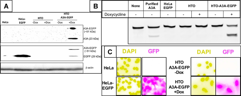
Figure 1. Characterization of a HeLa cell line inducible for A3A-EGFP.
HeLa-derived cell lines were induced for 12 hr A3A-EGFP expression using doxycycline (Dox) and analyzed for protein expression (A), cytosine deamination activity (B) or GFP fluorescence (C). The different cell lines used, HeLa-EGFP, HTO and HTO-A3A-EGFP are described in the text. A. Western blot analysis of A3A-EGFP (upper band) and EGFP (lower band) using anti-A3A/B or anti-GFP antibodies with β-actin as the loading control. A3A-EGFP is only detectable in induced HeLa-TO A3A-EGFP cell lysate. B. Cytosine deamination activity in lysates of different cell lines with or without induction. A 6FAM-labeled oligomer containing a single cytosine was sequentially treated with cell extracts, Ung and NaOH at 95°C. The shorter cleaved product was separated from the substrate on a denaturing gel. The oligomer was also incubated with partially purified A3A to confirm the position of the product on the gel. C. The cells were stained with DAPI and visualized using fluorescence. The colors in the all the images were inverted to create greater contrast between different colors.

