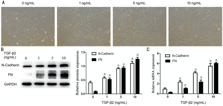
Figure 4. TGF-β2 induce EMT in RPE cells.
RPE cells were exposed to three different concentrations (1, 5, 10 ng/mL) of TGF-β2 for 48h and 0 ng/mL TGF-β2 is a blank control group. A: The cell morphological appearance was analyzed by a phase-contrast microscope at 100× magnification; B: Western blotting showed TGF-β2 could induce N-Cadherin and FN proteins expression with obvious dose-dependence. GAPDH was selected for internal reference (aP<0.05). C: qRT-PCR analysis showed N-Cadherin and FN mRNA expression increases with the TGF-β2 concentration. GAPDH was selected for internal reference (aP<0.05). Performed all experiments in triplicate.

