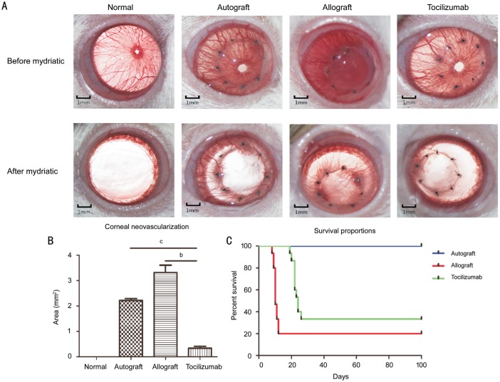
Figure 2. Clinical observation of corneal graft after corneal transplantation.
A: The appearance of corneal grafts after transplantation. Fourteen days after transplantation, the opacity, area of neovascularization and edema were observed under the microscopy before and after mydriasis. B: Corneal neovascularization area in each group after mydriasis (n=15). bP<0.01, cP<0.001. C: Survival percentages after surgery. On postoperative day 100, the corneal graft and the rejection score were recorded for drawing the survival curves (n=15).

