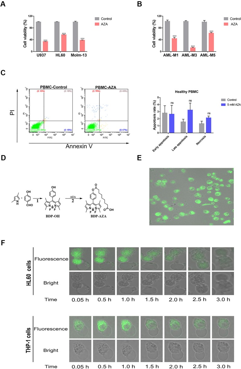
Figure 1.
Azelaic acid (AZA) inhibits acute myeloid leukemia (AML) cell proliferation. (A) The U937, HL60, and Molm-13 cells were treated with AZA at concentration of 5.0 mM for 48 h. Cell viability was measured by the CCK-8 method. (B) AML patient cells were isolated and then treated with 5 mM AZA for 48 h. Cell viability was measured by the CCK-8 method. (C) Peripheral mononuclear cells (PBMC) were treated with 5 mM AZA and then stained with Annexin V/PI. The apoptotic rate was measured by flow cytometry analysis. (D) The synthetic method of BDP-AZA. (E) HL60 cells were cultured in confocal dish for 24 h and 1 mM BDP-AZA was added to the medium. Thereafter, cell membrane was observed to turn green with confocal microscope under the excitation of blue light. (F) HL60 and THP-1 cells began to develop swelling and cytoplasmic vacuolization under fluorescence confocal microscopy after AZA treatment. A total of three independent experiments were performed, ***P < 0.001. ns, no significance.

