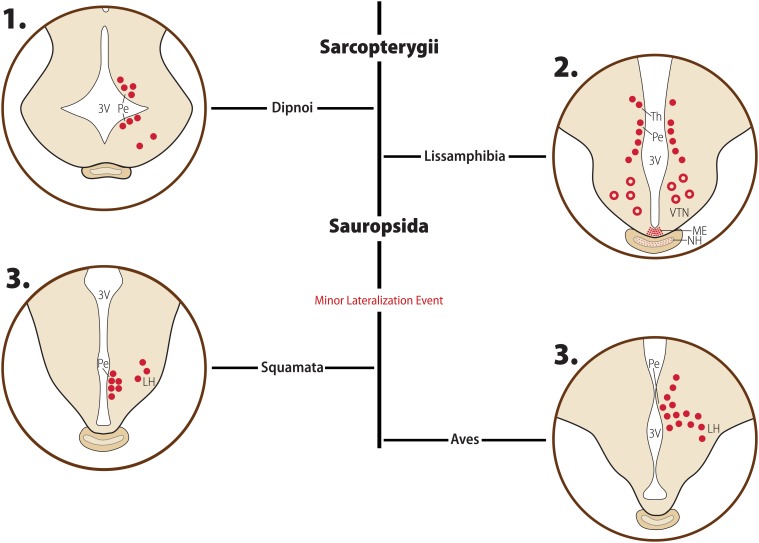
FIGURE 5.
Diagrammatic representation of the main morphological features of the MCH distribution in non-Mammalian Sarcopterygians. Major clades are indicated in bold font, while clade-wide events are represented in red. The diagrams for each clade are not meant to represent a single species or a single hypothalamic level, but rather a visual summary of what has been described for animals belonging to that clade. Full red circles represent MCH neurons, open red circles represent inconsistent information in the literature about MCH-ir neurons in the area. Fiber plexuses are represented by red dots, and inconsistent data about innervation is represented by red stars. 1. Lungfish (Dipnoi) have a distribution of cells that closely resembles that of Elasmobranchs and early Actinopterygians, with neurons concentrated around the ventricle (Pe), and only scattered cells found in lateral areas. 2. In Lissamphibians, neurons are still concentrated in the dorsomedial hypothalamus, but these cells are not as close to the ventricle as in other species, and some of the extend dorsally toward the ventral thalamus (Th). Fibers are observed in the Lissamphibian median eminence (ME). There is no consensus about the presence of MCH neurons in the ventral tuberal nucleus (VTN), represented by red open circles, or about the innervation of the neurohypophysis (NH), represented by red stars. 3. In Sauropsids, a lateral migration event occurred, with some neurons found forming an arc shaped in the lateral hypothalamus (LH). These neurons do not surpass the number of periventricular neurons, however, making this a minor lateral migration event. Based on data from [opetwcite]B137,B35[clotwcite]Vallarino et al. (1998); Cardot et al. (1994), [opetwcite]B36,B58[clotwcite]Cardot et al. (1999); Francis and Baker (1995), and Lázár et al. (2002).

