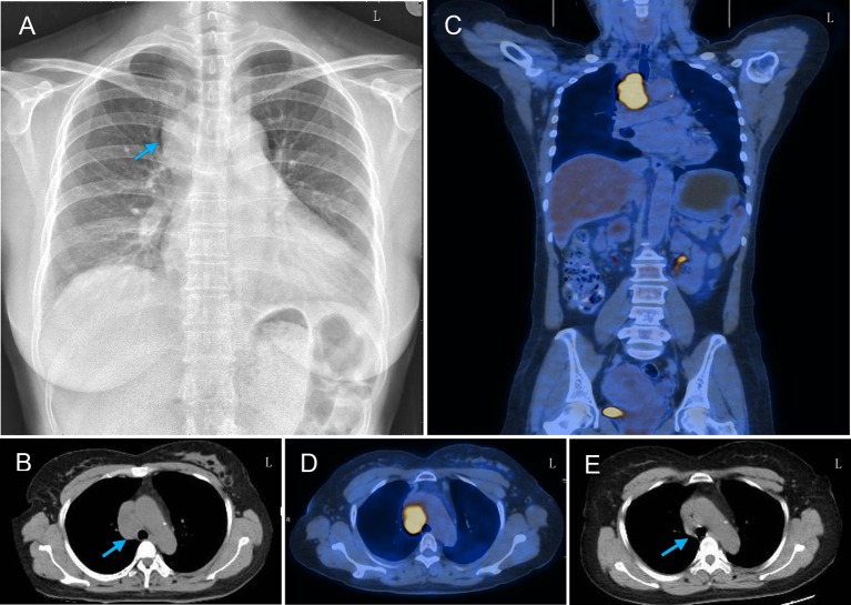
Figure 2.
Thoracic radiograph and PET-CT findings of the patient. (A) Chest X-ray showed a round soft tissue mass (arrow) in the upper-mid mediastinum and infiltrating right lung field. (B) Thoracic CT showed a round soft tissue mass (arrow) located in the upper-mid mediastinum before the arteroae aorta. (C,D) PET-CT scan showed increased uptake of fluorodeoxyglucose signal in this mass axial and coronal plane images. (E) Repeated thorax CT showed the resolution of the mediastinal mass after chemotherapy.

