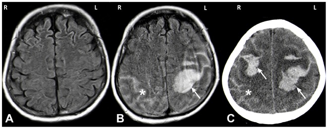
Figure 1.

MRI (A,B) and CT (C) images. MRI imaging on day of admission after a first seizure was unremarkable (A: axial FLAIR). MRI on day 5 showed a spontaneous left hemispheric ICH with a subarachnoid component (SAB) (B: axial FLAIR; white arrow: ICH, asterisk: SAB). One day later the patient deteriorated again and CT imaging showed a right sided sICH and edema of the left hemisphere (C: axial CT; white arrow: ICH, asterisk: SAB).
