
Figure 2.
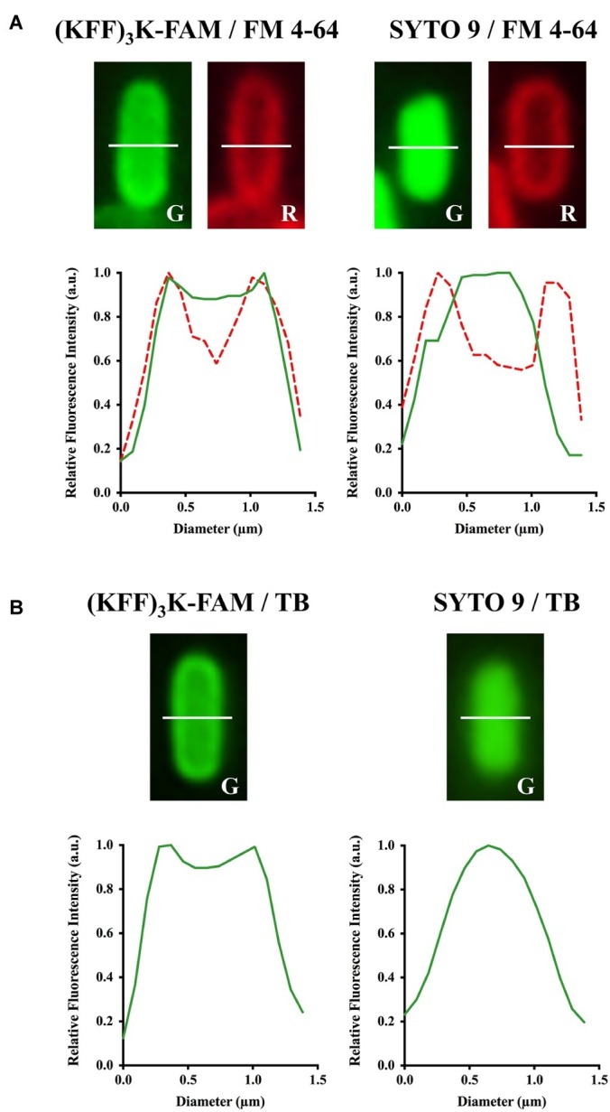
Confirmation of (KFF)3K-FAM permeation into the cytoplasm of E. coli. (KFF)3K-FAM or SYTO 9 treated cells were stained with (A) FM 4–64 or (B) Trypan Blue (TB). (KFF)3K-FAM permeation and dye staining are as performed in Figure 1. E. coli cells stained with SYTO 9 and either FM 4–64 or TB were used as controls. Single cells were randomly selected from fluorescence microscopic images and fluorescence values from the cross section (white dotted line) of the respective staining combinations were extracted to provide quantitative representations of internalized (KFF)3K-FAM. The fluorescence values for TB stained cells in the red fluorescence channel were equivalent to the background, thus excluded from the current analysis.