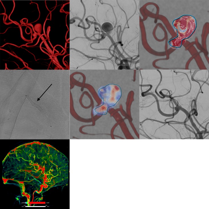
Figure 1.
Example of flow diverter stent treatment in the right peripheral middle cerebral artery (M2–3 segment) of a patient with two closely adjacent saccular aneurysms. The upper row (from left to right) shows the three-dimensional angiogram, corresponding conventional digital subtraction angiography (DSA) image in working projection, and intra-aneurysmal flow quantification (employing two-dimensional vector-based imaging), revealing long-lasting turbulent vortical flow in both aneurysmal compartments. The middle row shows the implanted Silk Vista Baby (2.25 mm x 15 mm, arrow), the immediate reduction of aneurysmal influx in vector-based imaging (reduced influx and decreased flow velocity in comparison with the pretreatment image), and the strongly decreased filling of both aneurysms in the conventional DSA image. The bottom row shows maintained normal perfusion of the right hemisphere including the parenchyma from the treated vessels.

