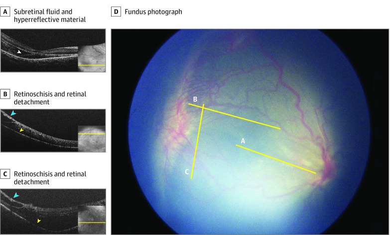
Figure 2. Handheld Optical Coherence Tomography (OCT) Images and Fundus Photograph of a Right Eye With Stage 4B Retinopathy of Prematurity With Both Retinoschisis and Retinal Detachment.
A, An OCT B-scan through the fovea shows subretinal fluid and subretinal hyperreflective material (white arrowhead). B and C, OCT B-scans through the retinal midperiphery in the area of perceived retinal detachment on indirect ophthalmoscopy shows both retinoschisis (blue arrowheads) and retinal detachment (yellow arrowheads). The yellow lines mark the approximate location of the OCT B-scans on OCT retina view (insets) and fundus photograph (D).

