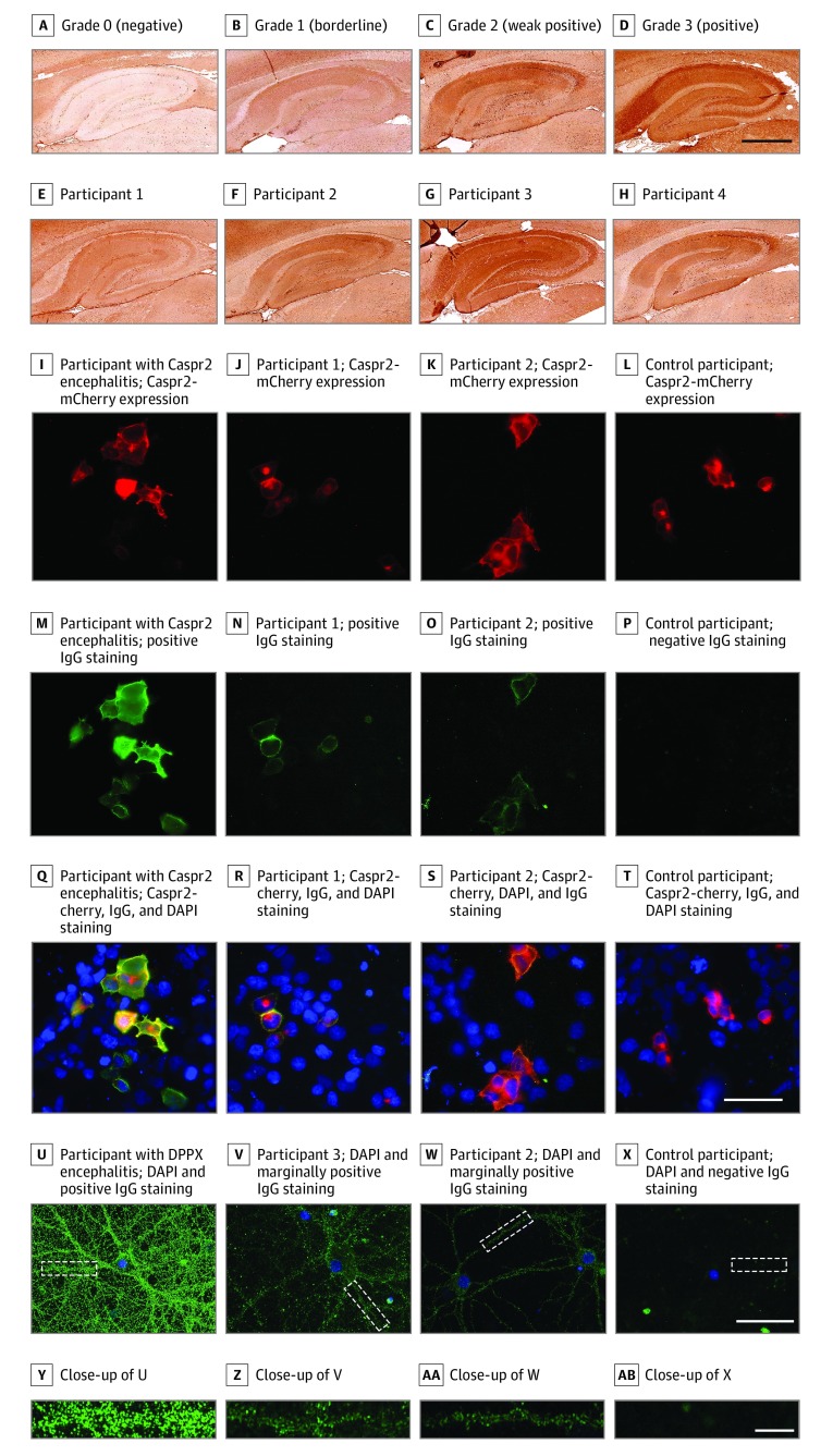
Figure. Autoantibody Detection Strategy and Representative Results.
A-H, Representative results of rat-brain immunohistochemistry testing using human sera. Autoantibody reactivity was shown by using a biotinylated antihuman IgG-specific secondary antibody, streptavidin-conjugated peroxidase and diaminobenzidine. The grading of rat-brain immunohistochemistry was based on 4 staining intensities of the hippocampus (A, grade 0, from a healthy control participant; B, grade 1, and C, grade 2, from the sera of the affected cohort; and D, grade 3, from the serum of an patient with autoimmune encephalitis and anti–contactin-associated protein-like 2 [Caspr2] antibodies). E-H, Samples that were also reactive on cell-based assays or neuronal staining. Scale bar = 500 μm (D). I-T, Results from live cell-based assays for Caspr2. Human sera were incubated on live human embryonic kidney 293 cells transfected with Caspr2 (with mCherry tag [red], I-L) and stained with antihuman IgG Alexa-488 (green). The presented results include a positive result from a participant with anti-Caspr2 encephalitis (M and Q; the same serum as in D) and individuals with psychosis (N and R) or from the control group (O and S) and a negative sample from a control participant (P and T). Nuclei were stained with 4′,6-diamidino-2-phenylindole (DAPI; blue). Q-T, Images show mCherry, DAPI, and positive IgG staining. Scale bar = 50 μm (T). U-X, Staining on rat hippocampal primary live neurons. Neurons were incubated with human serum samples and subsequently with antihuman IgG Alexa-488 and DAPI. Each image represents 1 or 2 neurons (scale bar = 50 μm [X]), and underneath, the zoomed-in image of the area identified with a white outline (scale bar = 10 μm [AB]). U, A positive result is represented by serum from a patient with encephalitis and autoantibodies against dipeptidyl-peptidase-like protein-6 (DPPX); V, sera of a patient with schizophrenia (the same individual as in G) and an individual from the control cohort (the same individual as in H), both tested with low reactivity on neurons, and the control participant with a negative result was represented by serum from a healthy individual. All sera were diluted 1:50. Nuclei were stained with DAPI.

