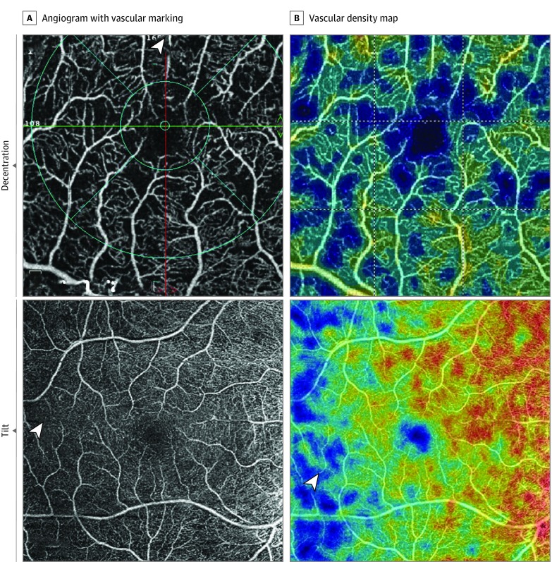
Figure 2. Technical-Related Artifacts.
Decentration artifact (top panels): Fovea is shifted superiorly with loss of more than 10% of inner subfield as shown by the arrowhead. Tilt artifact (bottom panels): Symmetric decrease in vascular density on temporal half of the angiogram and map (arrowheads) is shown.

