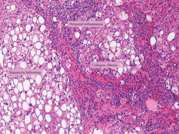
Fig. 1.
Liver biopsy specimen (Masson's trichrome stain, × 100) showing macrovesicular steatosis, diffuse hepatocyte ballooning (HB) and rare Mallory's hyaline bodies (MB). Periportal inflammation and fibrosis suggest cirrhotic changes in liver parenchyma. These histopathological changes are consistent with advanced NASH.

