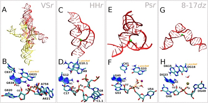
Figure 2.
Structures and active sites of VSr, HHr, Psr, and 8-17dz. Shown are the crystal structures and representative active sites in their active protonation state (e.g., general base guanine deprotonated at the N1 position) and conformational state derived from MD simulations in solution. (A, B) VSr dimer crystal structure12 (PDB ID 5V3I) and MD active state; (C, D) HHr crystal structure13 (PDB ID 2OEU) and MD active state;8 (E, F) Psr crystal structure14 (PDB ID 5K7C) and MD active state;9 (G, H) 8-17dz crystal structure15 (PDB ID 5XM8) and MD active state.10

