Fig. 1.

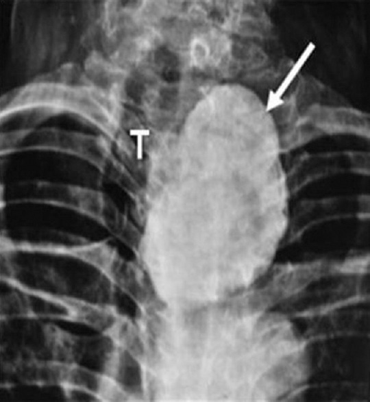
X-ray of the neck (anteroposterior view) showing a large calcified mass (arrow) displacing the trachea (T) towards the right side.

X-ray of the neck (anteroposterior view) showing a large calcified mass (arrow) displacing the trachea (T) towards the right side.