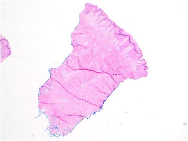
Figure 2.

At 2× magnification, histopathological analysis reveals haphazardly arranged smooth muscle bundles throughout the reticular dermis, sclerotic intervening dermis and small collapsed blood vessels with mild perivascular lymphocytic infiltrate.
