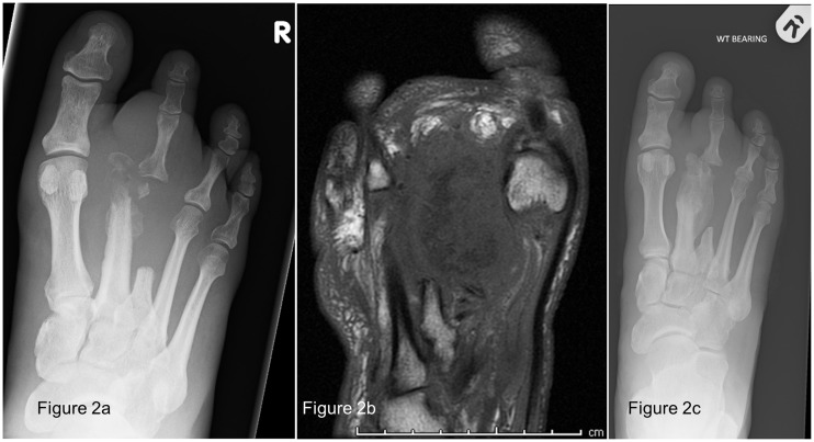
Figure 2.
(a) Radiograph of the right foot of Case 2 showing new bone destruction centred on the neck of the second metatarsal with dislocation of the second metatarsophalangeal joint, in keeping with osteomyelitis, (b) Magnetic resonance imaging of the right foot of Case 2 showing extensive osteomyelitis within the second metatarsal with bone fragmentation of the metatarsal head and involvement of the metatarsophalangeal joint, as well as extensive periosseous inflammatory phlegmon formation around the second metatarsal shaft and (c) radiograph of the right foot of Case 2 showing ongoing exuberant bone formation, bony erosion and periosteal reaction of the right second metatarsal.

