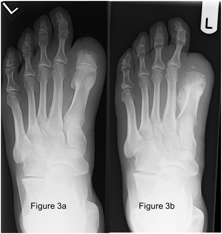
Figure 3.
(a) Radiograph of the left foot of Case 3 showing new ossification centre at the site of the stump, with remodelling of the residual proximal phalanx of the first toe and (b) radiograph of the left foot of Case 3 showing new callous formation and periosseous soft tissue swelling at the first metatarsal, with new erosions at the medial aspect and a periosteal reaction at the lateral aspect.

