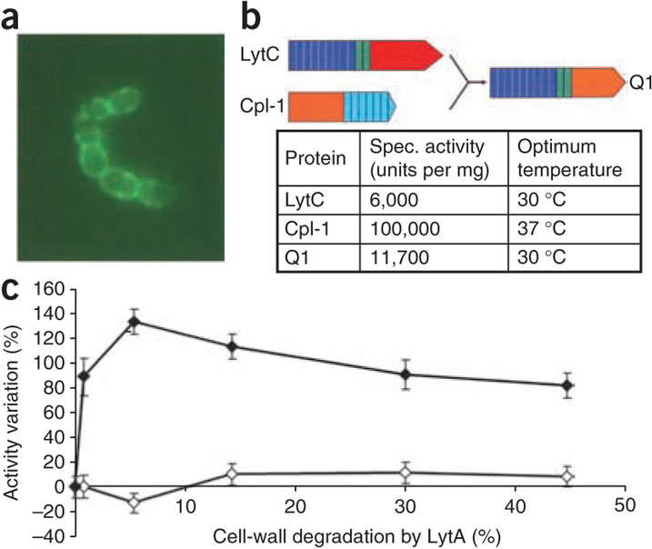
Figure 2.
Localization of GFP-LytC, properties of Q1 chimeric protein, and differential behavior between LytC and Cpl-1. (a) Subcellular localization of LytC by fluorescence image of the GFP-LytC fusion protein added to the pneumococcal culture. (b) Schematic representation of the Q1 chimeric protein. The modular construction of the parental proteins is represented by different colors: red and orange for catalytic modules of LytC and Cpl-1, respectively; deep blue and green for repeats of the choline-binding motifs of LytC, and light blue for repeats of the choline-binding motifs of Cpl-1. The relevant enzymatic properties of the parental and chimeric proteins are presented. (c) Increase of in vitro LytC activity by the pretreatment of pneumococcal cell walls with small amounts of LytA added to cell walls before the action of LytC or Cpl-1.

