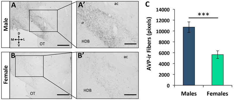
Figure 1. Sex difference in vasopressin immunoreactive (AVP-ir) fiber density in the ventral pallidum (VP).

(A-B) Low (A, B; 10X; scale bar = 600μm) and high magnification (A’, B’; 20X; scale bar = 200 μm) photomicrographs depict a high density of AVP-ir fibers present in the VP [+0.60mm from Bregma according to Paxinos and Watson (2007)] of adult male rats (A-A’), compared to adult female rats (B-B’). (C) Quantification of AVP-ir fiber density in the VP (averaged over three 30-μm sections per rat) shows higher AVP-ir fiber density in adult male rats (n=7) compared to adult female rats (n=7). Data are represented as the mean ± SEM, two-tailed t-test; ***p<0.005. ac = anterior commissure; OT = olfactory tubercle; HDB = horizontal diagonal band of Broca.
