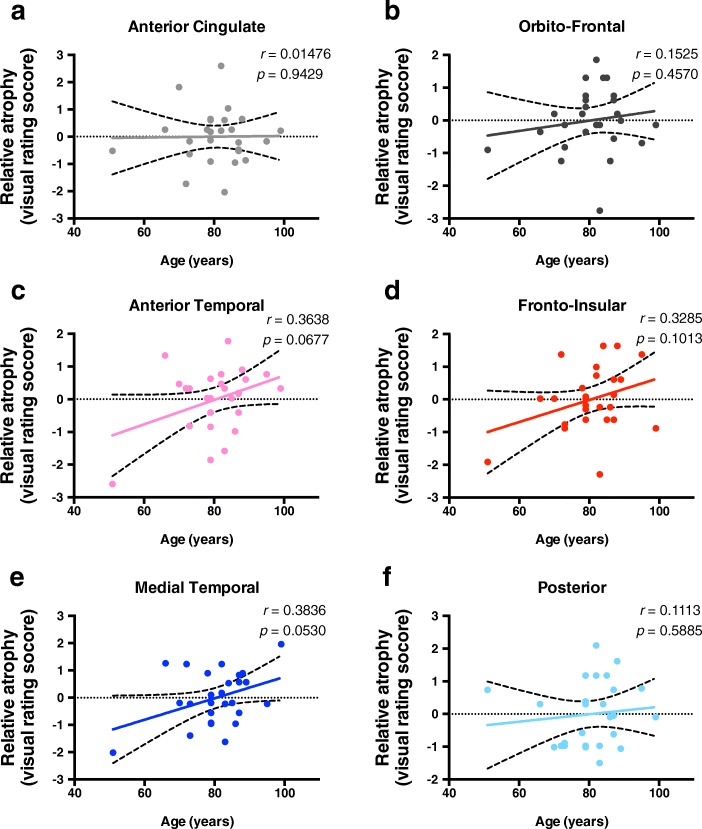
Fig. 3.
Anterior and Medial temporal lobe atrophy increases with increasing age. The values represented are residuals corrected for Braak staging, with Pearson correlation analysis between age and atrophy of different brain regions based on a previously validated imaging rating scale. The regions evaluated are a anterior cingulate, b orbito-frontal, c anterior temporal, d fronto-insular, e medial temporal and f posterior brain regions. Since statistical significance was considered as p < 0.05, no significant correlations were found, although both anterior temporal (r = 0.3638, p = 0.0677), and medial temporal (r = 0.3836, p = 0.053) regions were found to be close to this statistical threshold

