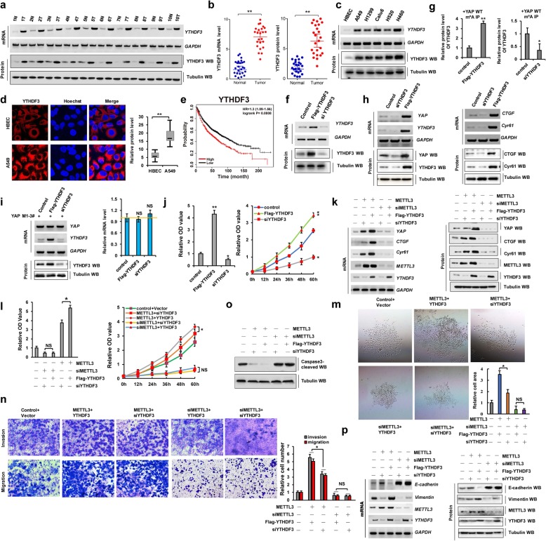
Fig. 2.
YTHDF3 recognizes m6A modification and promotes cellular growth and migration via YAP upregulation. a–d RT-PCR, western blot and Immunofluorescent staining assay indicated that the mRNA and protein levels of YTHDF3 were higher in human lung cancer tissues (a, b) and cell lines (c, d) compared with their normal adjacent lung tissues and control cell, HBEC. e Kaplan–Meier overall survival (OS) curves of YTHDF3 (p = 0.0008 by log-rank test for significance) of human lung cancers. f The expressions of YTHDF3 were analyzed by RT-PCR and western blot. g The interaction between YTHDF3 and YAP mRNA was analyzed by RIP from A549 cells immunoprecipitated with m6A antibody. h The expressions of YAP and its target genes, CTGF and Cyr61, were analyzed by RT-PCR and western blot. i The relative mRNA level of YAP was analyzed by RT-PCR in A549 cells with transfection with indicated genes. j The cellular growth was analyzed by CCK8 assay in A549 cells with transfection with Flag-YTHDF3 or siYTHDF3, respectively. k–o A549 cells were respectively correspondent co-transfection with Ov/si-METTL3 and Ov/si-YTHDF3 as the indication. k The expressions of METTL3, YTHDF3, YAP and its target genes, CTGF and Cyr61, were analyzed by RT-PCR and western blot. l The cellular proliferation and growth were analyzed by CCK8 assay. m Colony formation ability was analyzed by colony formation assay. n The cellular invasion and migration growths were analyzed by transwell assay. o The protein level of cleaved Caspase-3 was analyzed by western blot. p The mRNA and protein levels of E-cadherin and vimentin were analyzed by RT-PCR and western blot assay. Results were presented as mean ± SD of three independent experiments. *P < 0.05 or **P < 0.01 indicates a significant difference between the indicated groups. NS, not significant

