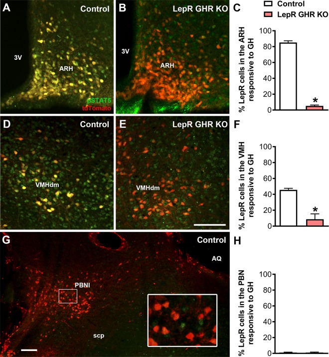
Figure 1.
GH-responsive neurons in the hypothalamus of control and LepR GHR KO mice. LepR-expressing cells (red) and pSTAT5 (green) 90 min after an intraperitoneal injection of porcine GH (20 µg/g) in LepR-IRES-Cre::LSL-tdTomato mice (control) and LepR GHR KO::LSL-tdTomato mice. Yellow represents double-labeled cells. The inset represents a higher magnification photomicrograph of the selected area. 3V, third ventricle; AQ, cerebral aqueduct; scp, superior cerebellar peduncle; VMHdm, dorsomedial subdivision of the VMH. Scale bars, 100 µm. *P < 0.05 (unpaired, 2-tailed Student’s t test).

