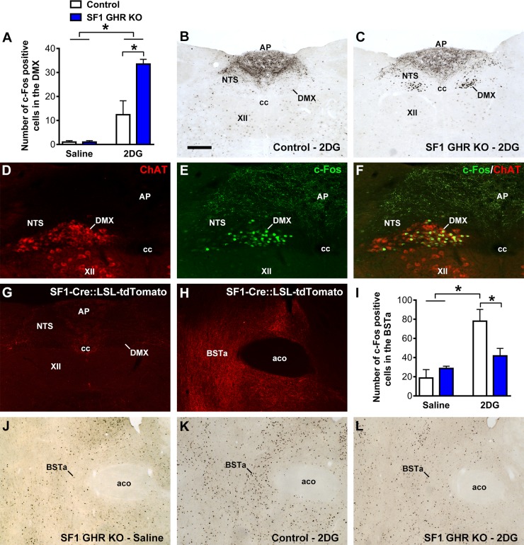
Figure 6.
GHR ablation in SF1 cells affects the activity of the neurocircuit that regulates the PNS. A) Number of c-Fos–positive cells in the DMX 2 h after intraperitoneal injections of saline or 2DG {interaction between 2DG effect and GHR ablation [F(1, 11) = 6.039, P = 0.0318]; n = 3–5}. B, C) Photomicrographs showing 2DG-induced c-Fos expression in the DMX of control and SF1 GHR KO mice. D–F) Epifluorescence photomicrographs showing the colocalization between ChAT (red) and c-Fos (green) in the DMX of SF1 GHR KO mice. G, H) Epifluorescence photomicrographs showing the distribution of SF1/tdTomato axons in the vagus complex (G) or the BSTa (H) using the in SF1-Cre::LSL-tdTomato reporter mouse. I) Number of c-Fos–positive cells in the BSTa 2 h after intraperitoneal injections of saline or 2DG {interaction between 2DG effect and GHR ablation [F(1, 12) = 5.079, P = 0.0437]; n = 3–5}. J–L) Photomicrographs showing 2DG-induced c-Fos expression in the BSTa of saline-treated SF1 GHR KO (J), 2DG-treated control (K), and 2DG-treated SF1 GHR KO mice (L). Aco, anterior commissure; AP, area postrema; cc, central canal of the spinal cord; NTS, nucleus of the solitary tract; XII, hypoglossal nucleus. Values are means ± sem. Scale bars: 200 µm (B, C); 100 µm (D–F); 400 µm (G); 200 µm (H, J–L). *P < 0.05 (Bonferroni’s multiple comparisons test).

