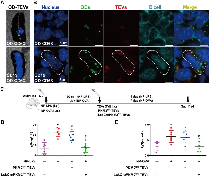
Figure 6.
Nanomaterial packaging with TEVs modulates B-cell IgG production in vivo. A, B) Animals administered tail-vein injections of the nanocomposites CD19-QD-CD63-TEVs or QD-CD63-TEVs (50 μg of TEVs/mouse) in C57BL/6J mice. Representative micrographs of the specificity of the CD19-QD-CD63-TEVs (TEVs labeled with PKH26) in mouse spleen tissues (A) and splenic B220-positive B cells (B). C) Schematic flowchart of C57BL/6J mice that were injected intraperitoneally with 100 ng of NP-LPS or 100 ng of NP-OVA antigen; after 30 min or 1 d, mice were given tail-vein injections of CD19-QD-CD63-TEVs (LckCrePKM2fl/fl-TEVs vs. PKM2fl/fl-TEVs). D) Plasma IgG levels induced by NP-LPS antigen were measured via ELISA. E) Plasma IgG levels induced by NP-OVA antigen were measured via ELISA; n ≥ 5, data are presented as means ± sd. *P < 0.05 vs. the control (CTL) group, #P < 0.05 vs. the NP-LPS + PKM2fl/fl-TEVs group (D) or NP-OVA + PKM2fl/fl-TEV group (E) (1-way ANOVA followed by Tukey’s test for multiple comparisons).

