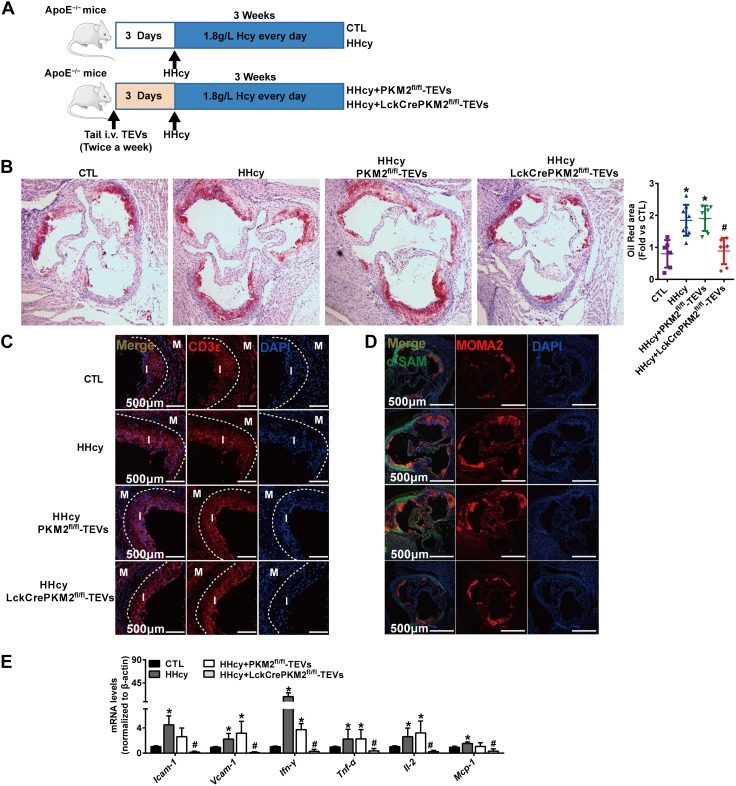
Figure 8.
Nanomaterial packaging with PKM2-null TEVs inhibits HHcy-accelerated atherosclerosis. A) Schematic flowchart of ApoE−/−mice tail-vein injected with CD19-QD-CD63-TEVs (LckCrePKM2fl/fl-TEVs vs. PKM2fl/fl-TEVs) twice per week; 3 d after the first injection, HHcy-accelerated atherosclerosis was induced by giving mice drinking water supplemented with 1.8 g/L Hcy. B) Oil Red O staining of aortic roots for quantitative lesion size (left panel). Quantification of the mean atherosclerotic lesion area is shown (right panel). C) Representative confocal images of T-cell infiltration (positive CD3 staining). D) Representative confocal micrographs of the infiltration of macrophages/macrophage [macrophages/monocytes antibody (MOMA2) staining positive] in lesion areas; there were no obvious changes in the α-SMA-stained area. E) The gene expression of Icam-1, Vcam-1, Ifn-γ, Tnf-α, Il-2, and CCL2 in thoracic aortas isolated from mice was measured via qPCR; n = 5–8, data are presented as means ± sd. *P < 0.05 vs. control (ctl) group;. #P < 0.05 vs. HHcy-PKM2fl/fl-TEVs group (1-way ANOVA followed by Tukey’s test for multiple comparisons).

