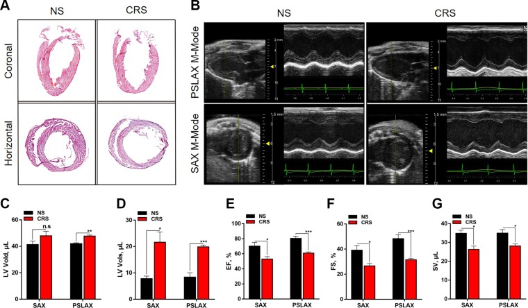
Figure 2.
Results of cardiac H-E staining and echocardiography of 10-wk-old CRS and NS mice. A) H-E staining of whole hearts sectioned along the coronal or horizontal planes. Compared with NS controls, the hearts of CRS mice showed LV chamber dilatation and wall thinning, suggesting that CRS induced cardiac remodeling and heart failure. B) Representative echocardiographic images under parasternal long axis (PSLAX) and short axis (SAX) M-mode. The images show LV chamber dilatation and decline of LV contraction. C–G) Measurements of echocardiographic parameters in CRS and NS mice: LV diastolic volume (LV Vold), microliters (C); systolic volume (LV Vols), microliters (D); ejection fraction (EF), percentage (E); fractional shortening (FS), percentage (F); and stroke volume (SV), microliters (G). N.s., not significant. *P < 0.05, **P < 0.01, ***P < 0.001 vs. NS group (n = 8 in NS group, n = 9 in CRS group).

