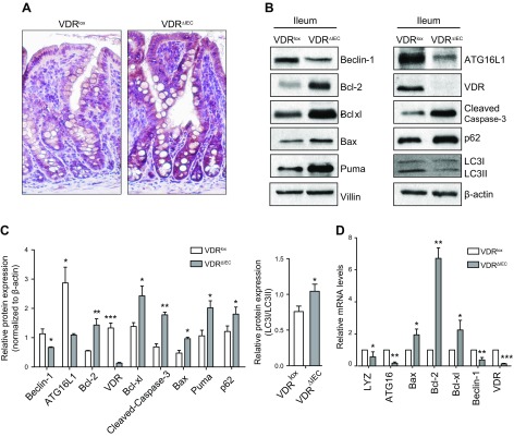
Figure 2.
Apoptosis- and autophagy-related genes were regulated by intestinal epithelial VDR. A) Enhanced cleaved caspase-3 in VDR∆IEC mice according to immunohistochemistry staining. Representative images of cleaved caspase-3 immunohistochemistry in small intestinal crypts from VDRlox and VDR∆IEC mice. B) Western blots for Beclin-1, Bcl-2, Bcl-xL, Bax, P62, LC3I/II, PUMA, and cleaved caspase-3 in the small intestines of VDR∆IEC and VDRlox mice. We found reduced levels of Beclin-1 and enhanced levels of Bcl-2, Bcl-xL, P62, and cleaved caspase-3. C) The intensity of gene expression in the intestinal tissues was analyzed by Western blotting, and the ratio of their expression to that of β-actin was calculated. D) Real-time PCR for Beclin-1, Bcl-2, Bcl-xL, Bax, LYZ, and ATG16L1 in the intestines of VDR∆IEC and VDRlox mice. LC3, autophagy-related proteins, light chain 3; LYZ, lysozyme. The data represent means ± sem; Student’s t test (n = 5 mice/group). *P < 0.05, **P < 0.01, ***P < 0.001.

