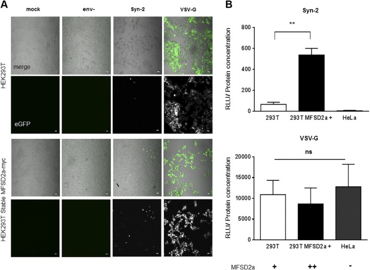
Figure 3.
Syn-2–pseudotyped viruses are infectious in MFSD2a-expressing cells. A) Confocal microscopy images of infected cells. Parental and stably MFSD2a-overexpressing HEK293T cells were mock-infected or infected with env-, Syn-2–, and VSV-G–pseudotyped viruses and GFP expression was next detected by confocal microscopy. Scale bar, 10 µm. B) Luciferase activities were measured from HEK293T and HeLa cells infected with luciferase-expressing Syn-2–pseudotyped viruses (top panel) or VSV-G–pseudotyped viruses (bottom panel). For each sample, luciferase activity was quantified 24 h postinfection (expressed as RLU) and normalized against protein concentration. Signs (+ and –) under the graphs indicate MFSD2a cellular expression level. These results are representative of 3 independent experiments. Ns, not significant **P < 0.01 (unpaired Student t test).

