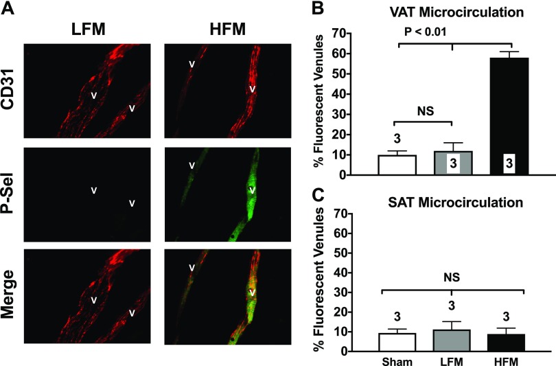
Figure 1.
A single HFM up-regulates P-selectin in the microcirculation of the mesenteric VAT. A) Photomicrographs show typical fluorescent staining for P-selectin (green staining; middle panel of right column) observed in the mesenteric VAT microcirculation of mice given a single HFM. Endothelial P-selectin was detected using a mAb that selectively recognizes surface-expressed P-selectin (green). Constitutively expressed platelet and endothelial cell adhesion molecule 1(PECAM-1) was also cross-linked (red stain) to confirm the vascular nature of the stained structures and to normalize expression levels of P-selectin. B, C) Specific secondary antibodies conjugated with fluorescein (P-selectin) and rhodamine (PECAM-1) were used. HFM acutely up-regulates P-selectin in the mesenteric VAT microcirculation (B) but not in the subcutaneous fat (SAT) microcirculation (C). Studies were performed 120 min after gavage administration of a liquid HFM to control C57BL/6J mice. Bar graphs show quantification of P-selectin staining in all groups of mice. Values are means ± se. Numbers at the bottom of bars indicate the number of mice studied in each group. Forty venules were counted in each mouse. NS, not significant. All images were obtained at ×200 magnification.

