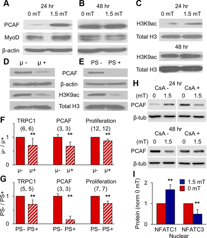
Figure 5.
Magnetic fields promote epigenetic MyoD. A, B) PEMF-modulated PCAF and MyoD protein levels at 24 (A) or 48 (B) h postplating. C) H3K9 histone acetylation levels after 1.5 mT PEMF exposure at 24 (top) or 48 (bottom) h postplating. D, E) PCAF protein and H3K9 acetylation levels after growth of myoblasts within a μ-metal box (D) or in the presence of PS (PS; 1%; for 72 h (E). F, G) Protein levels of TRPC1 and PCAF and proliferation after 72-h growth in a µ-metal box (F, µ+) or in the presence of PS (G, PS+) relative to controls (µ−, PS−); (n/condition). H) Effects of CsA (2 μM) over PEMF-modulated PCAF expression at 24 or 48 h. I) Nuclear and cytoplasmic distribution of NFATC1 and NFATC3 following 1.5 mT exposure (blue) at 25 h postplating (see also Supplemental Fig. S8); n = 6/condition. Data represent the means of n experiments ± sd), each derived from the means of triplicates. All PEMF exposures were 10 min. **P < 0.01 with regard to relative control.

