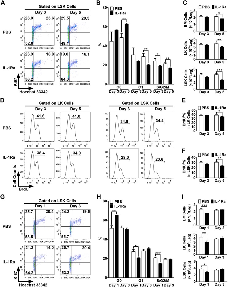
Figure 4.
rhIL-1Ra induced transient and reversible HS/PC quiescence in vivo. A–C) BALB/c mice received daily injections of either PBS or IL-1Ra (3 mg/kg) over 5 d. BMCs were harvested and analyzed at d 3 and 5. Levels of Ki67 and Hoechst 33342 staining were measured in LSK cells by flow cytometry. Representative dot plots (A) and mean percentages (B) of the LSK cells in G0, G1, and S/G2/M phases and the numbers of total BM, LK, and LSK cells per leg (C) are shown. D–F) The mice were treated with PBS or rhIL-1Ra as above and injected with BrdU (100 mg/kg) before euthanasia. BrdU incorporation was measured in LK and LSK cells by flow cytometry. Representative histograms (D) and mean percentages (E, F) of BrdU+ LK (E) and LSK (F) cells are shown. G–I) BALB/c mice received daily injections of either PBS or IL-1Ra (3 mg/kg) for 5 d. BMCs were harvested and analyzed at d 1 and 3 after the final injections (1 or 3 d recovery). Levels of Ki67 and Hoechst 33342 staining were measured in the LSK cells by flow cytometry. Representative dot plots (G) and mean percentages (H) of the LSK cells in G0, G1, and S/G2/M phases and the numbers of total BM, LK, and LSK cells per leg (I) are shown. All data are expressed as means ± sem (n = 3–6 mice/group at each time point). Statistical significance of the differences between groups was assessed with Student’s t test. *P < 0.05, **P < 0.01, ***P < 0.001.

