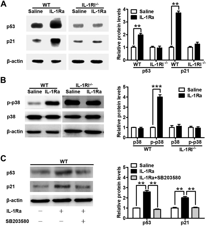
Figure 5.
rhIL-1Ra increased p53 and p21 protein levels by activating p38 MAPK in mouse BM. A) Western blot analysis of p53 and p21 in BM of WT and IL-1RI−/− mice after daily saline or rhIL-1Ra treatments for 5 d. The bar graph on the right indicates the means ± sd of the p53 and p21 levels. β-Actin was used as the housekeeping gene. The protein expression values were normalized to the saline control. **P < 0.01 (n = 3). B) Western blot analysis of phosphorylated p38 (p-p38) and p38 in BM of WT and IL-1RI−/−mice treated as above. β-Actin was used as the housekeeping gene. The protein expression values were normalized to the saline control. Data are means ± sd. ***P < 0.001 (n = 3). C) Western blot analysis of p53 and p21 in BM of WT mice treated daily with rhIL-1Ra (3 mg/kg) and the p38 inhibitor SB203580 (25 mg/kg) for 5 d. β-Actin was used as the housekeeping gene. The protein expression values were normalized to the saline controls. Results are means ± sd. **P < 0.01 (n = 3).

