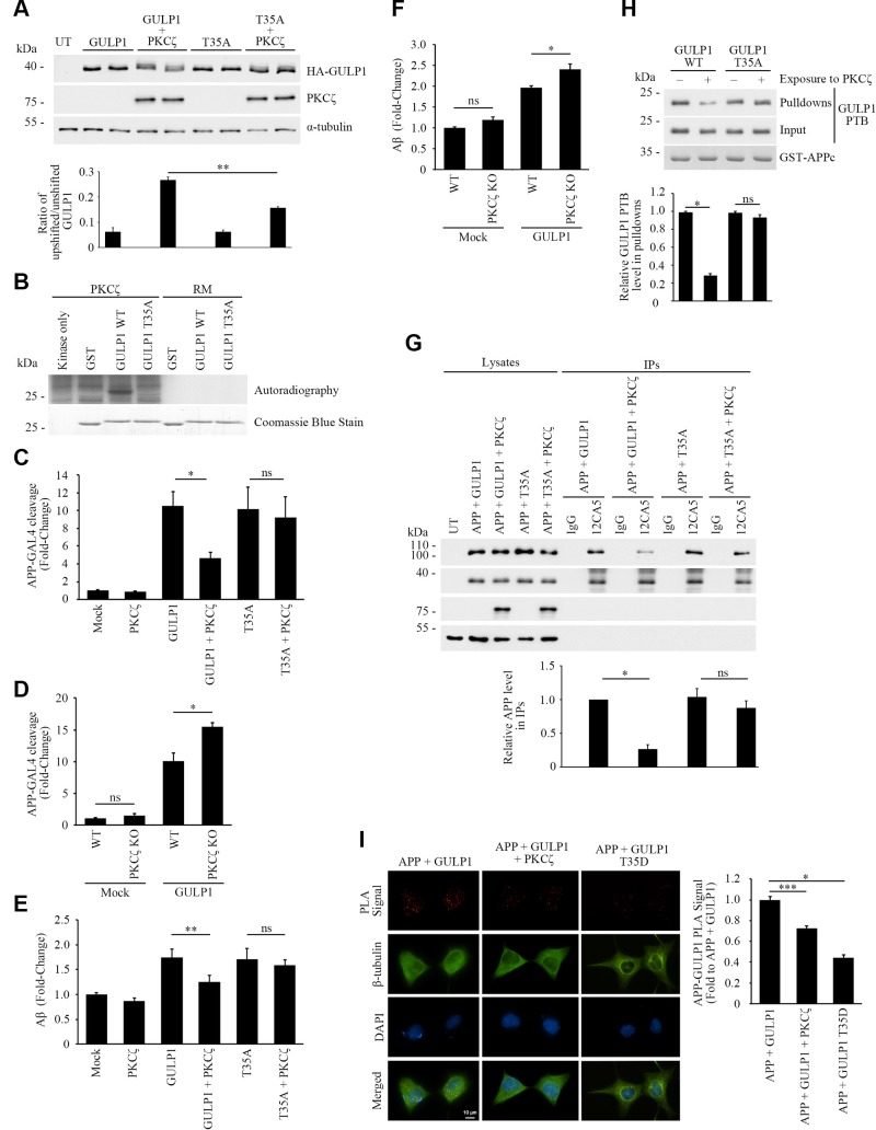
Figure 3.
PKCζ phosphorylates GULP1 T35 and reduces the effect of GULP1 on APP processing. A) HA-GULP1 and HA-GULP1 T35A were transfected to CHO cells together with or without PKCζ. Cell lysates were resolved on a high-resolution NuPAGE Bis-Tris Precast gel for determination of GULP1 migration pattern. Expression of FLAG-tagged PKCζ was confirmed by immunoblotting using anti-FLAG M2 antibody. α-Tubulin was detected by the anti–α-tubulin DM1A antibody as loading control. The amounts of unshifted and unshifted GULP1 were quantified by densitometry using Image Studio and their ratios in different transfected cells were illustrated. UT, untransfected (n = 5). **P < 0.01. B) Bacterially expressed GST-GULP126–43 (WT) or GST-GULP126–43 T35A (T35A) were incubated with PKCζ immunoprecipitated from transfected cell lysate together with (γ-[32P])-ATP for 30 min at 30°C. RM is the reaction mix only without kinase. Upper panel: autoradiograph; lower panel: Coomassie Blue staining PKCζ phosphorylates GULP1 at T35. C) HEK293 were cotransfected with APP-GAL4, pFR-Luc, and phRL-TK together with the indicated constructs. PKCζ reduces the effect of GULP1-mediated APP-GAL4 cleavage via phosphorylation of T35. Ns, not significant (n = 5). Results are means ± sd. *P < 0.001 compared with GULP1 transfected cells. D) WT or PKCζ knockout (KO) HEK293 cells were cotransfected with APP-GAL4, pFR-Luc, and phRL-TK together with the indicated constructs. GULP1-mediated APP-GAL4 cleavage was enhanced in PKCζ KO cells. Ns, not significant (n = 5). *P < 0.001. E) Cells were cotransfected with APP and the indicated constructs, and the level of secreted Aβ40 was assayed. PKCζ suppresses GULP1-enhanced Aβ40 liberation through phosphorylation of T35 (n = 5). Results are means ± sd. **P < 0.01 compared with GULP1 transfected cells. F) WT or PKCζ KO HEK293 cells were cotransfected with APP and the indicated constructs, and the level of secreted Aβ40 was assayed. GULP1-mediated Aβ40 secretion was increased in PKCζ knockout cells (n = 5). Results are means ± sd. *P < 0.001. G) PKCζ reduces GULP1-APP interaction in coimmunoprecipitation assay. Coimmunoprecipitation was performed from cells transfected with APP + HA-GULP1, APP + HA-GULP1 + PKCζ, APP + HA-GULP1 T35A, or APP + HA-GULP1 T35A + PKCζ using a mouse anti-HA antibody 12CA5. APP and HA-GULP1 in the immunoprecipitates (IPs) were detected by a rabbit anti-APP and 12CA5; n = 3. *P < 0.001. H) PKCζ reduces GULP1-APP interaction in vitro. Recombinant GULP1 PTB and GULP1 PTB T35A were phosphorylated in vitro by PKCζ. GST-APPc was used as bait to pull down the recombinant proteins. The amount of GULP1 PTB and GST-APPc was revealed by a rat anti-GULP1 and a rabbit anti-APP, respectively; n = 3. Results are means ± sd. *P < 0.001. I) PKCζ reduces GULP1-APP interaction in PLA. Cells were transfected with APP + GULP1, APP + GULP1 + PKCζ, or APP + GULP1 T35D. Fewer PLA signals were observed in APP + GULP1+ PKCζ and APP + GULP1 T35D cotransfected cells. β-Tubulin and DAPI were used as morphology and nucleus markers, respectively. Representative images are shown. Bar chart shows relative PLA signal (fold to APP + GULP1). Data were obtained from at least 60 cells per transfection and the experiments were repeated 3 times. Error bars = sem. *P < 0.001, ***P < 0.05.

