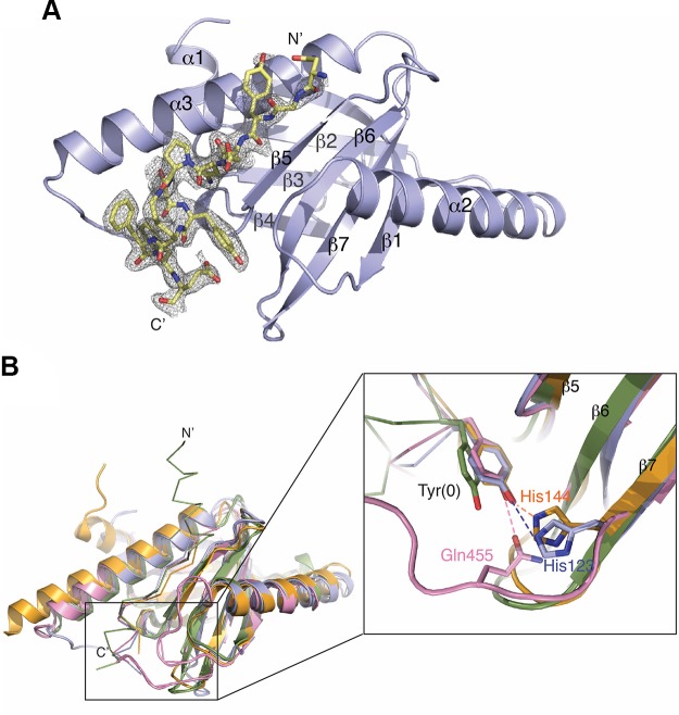
Figure 6.
Crystal structure of GULP1 PTB in complex with APP peptide. A) Overall structure of APP peptide bound to GULP1 PTB. Fo-Fc electron density map (contoured at 3σ) revealed large positive peaks and clearly indicates the presence of the APP peptide. B) Comparison of the overall structure of GULP1 PTB-APP structure and other PTB-APP complexes. GULP1 PTB-APP peptide complex (blue) is overlaid with Dab2 PTB-APP peptide complex [orange, PDB code: 1M7E], FE65 PTB-APP peptide complex (green, PDB code 3DXD), and X11 PTB-APP peptide complex (pink, PDB code: 1X11). The inset shows the side-chains of Tyr(0) in different complexes, except that of the FE65-APP peptide complex, interact with the PTBs in similar conformation. The side-chain of Tyr(0) of the APP peptide in the X11 PTB-APP complex structure is stabilized by Q455 from the β6-β7 loop (pink). On the other hand, Tyr(0) in the GULP1 PTB-APP and Dab2 PTB-APP complex structures interacts with H144 and H123 of the β7 strand of GULP1 (blue) and Dab2 (orange) respectively.

