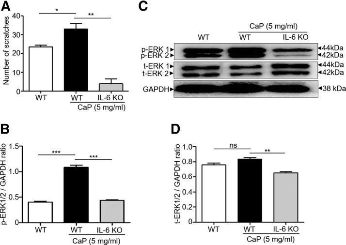
Figure 2.
The essential role of IL-6 in the CaP-induced elevation of p-ERK in DRG. A) Bar graphs show the number of scratches postinjection of CaP (0 or 5 mg/ml) in WT and IL-6 KO mice. B) Western blot analyses of p-ERK 1/2 and GAPDH in DRG of WT and IL-6 KO mice. C, D) The ratios of intensities of p-ERK 1/2 (C) and t-ERK 1/2 (D) relative to GAPDH were illustrated. Four mice per group were used for this experiment. The mean ± se for 3 separate experiments was calculated. *P < 0.05, **P < 0.01, ***P < 0.001.

