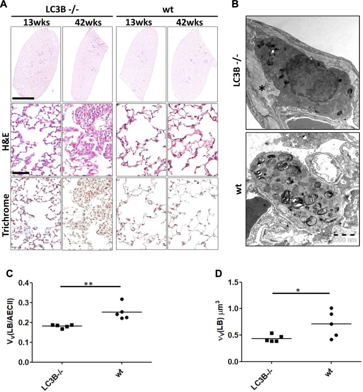
Figure 1.
Decreased airspaces and diameter of distal air spaces in aged LC3B−/− mice. A) Representative H&E staining of complete left lungs (upper panel) and higher magnification images of the H&E staining (middle panel) and trichrome staining (lower panel) of 13- and 42-wk-old LC3B−/− and WT mice. Scale for the whole lungs, 2 mm; scale for the higher magnification pictures, 100 μm; original magnification, ×200. B) Representative transmission electron microscopic images from 42-wk-old LC3B−/− and age-matched WT mice showing an example of smaller profiles of lamellar bodies in LC3B−/− compared with WT (primary original magnification, ×8900). C, D) Stereological data revealed a reduced volume fraction of lamellar bodies in AECII [VV(LB/AECII)] (C) combined with a reduced volume-weighted mean volume of lamellar bodies [νV(LB)] (D).

