Figure 9.
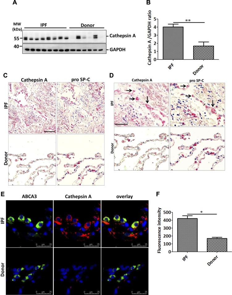
Increase in cathepsin A in AECII of IPF lungs. A, B) Western blot analysis of lung homogenates of patients with IPF and age-matched donors for cathepsin A and glyceraldehyde 3-phosphate dehydrogenase as loading control (A) followed by densitometric quantification for cathesin A (B). C, D) Serial paraffin sections from IPF and donor lungs were stained for cathepsin A and the AECII marker proSP-C and pictomicrographs were performed for the same areas from both types of staining. High magnification images (D) show several AECII cells also stained positively for cathepsin A, as indicated by the arrows. E) Immunofluorescence staining for the AECII lamellar body limiting membrane marker, ABCA3 (green) and cathepsin A (red). DAPI was used to stain nuclei in blue. F) Cathepsin A fluorescence signal intensity was quantified and depicted as a bar graph. Scale bars, 100 μm. Representative pictures are shown from ~10–15 patients with IPF and donors. **P < 0.01.

