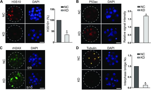
Figure 3.
Sin3a depletion results in cell proliferation failure and increased Trp53 acetylation. A) Immunostaining analyses of phosphorylation of H3S10, a hallmark for late G2 and mitosis, in mouse morulae (5–10 embryos/group; n = 3). Scale bar, 25 μm. B) Immunostaining analyses of Trp53K379 in morulae. The signal intensity of Trp53K79 was increased significantly (5–10 embryos/ group; n = 3. Scale bar, 25 μm). Nuclei were labeled with DAPI. C) Immunostaining analyses of γ-H2AX, an established marker for DNA damage in morulae. The experiment was replicated 3 times with 9 NC and 10 KD morulae analyzed. Scale bar, 25 μm. D) Immunocytochemical examination of α-tubulin, a marker for microtubule bridges, in morulae. Asterisk indicates microtubule bridge; n = 3; 5–10 embryos were analyzed/group each time. Scale bar, 25 μm.

