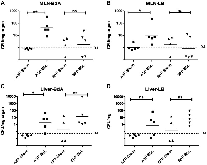
Figure 2.
Bacterial translocation (CFU) after BDL. A, B) CFU in MLNs of ASF-BDL and SPF-BDL mice 14 d after BDL, MLN-BdA (A) and MLN-LB (B). C, D) CFU in liver of ASF-BDL and SPF-BDL mice 14 d after (BDL). Geometric mean was calculated. Ns, not significant. Data are expressed as means ± sd; n = 4–5/group. *P < 0.05, **P < 0.005, ***P < 0.0005.

