Figure 3.
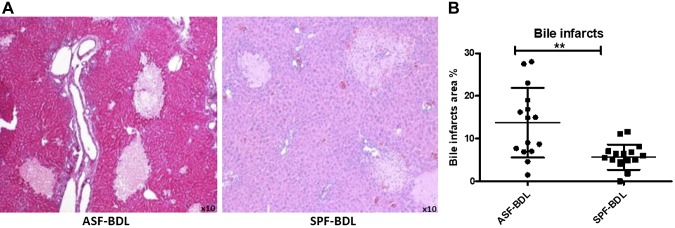
Evaluation of liver histology and bile infarcts area quantification. Histopathological examination of liver specimens and quantification demonstrated numerous bile infarcts in ASF-BDL and SPF-BDL mice. Determination of bile infarcts in the liver were assessed by conventional H&E staining (A) in ASF-BDL and SPF-BDL mice 14 d after BDL, then quantified (bile infarcts area percentage) using digital image analysis, MetaMorph (B) ASF-BDL and SPF-BDL mice 14 d after BDL. Data are expressed as means ± sd; n = 5 mice/group. *P < 0.05, **P < 0.005.

