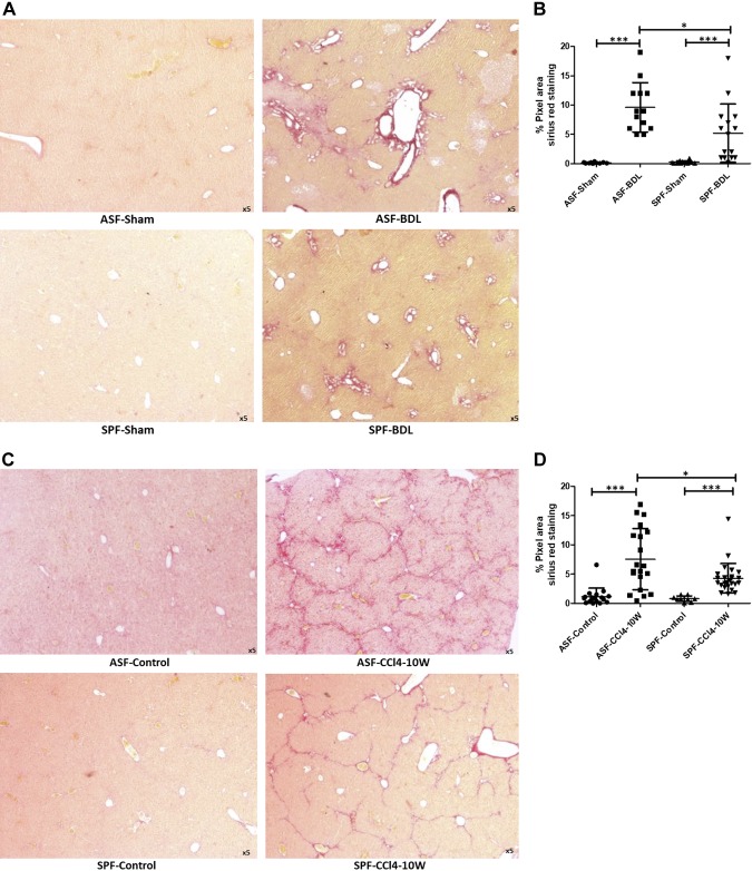
Figure 4.
Determination of fibrosis by collagen deposition within the liver sections in BDL- and CCl4-treated mice. Representative histologic images of livers stained with Sirius red to examine liver fibrosis ASF-sham vs. ASF-BDL (upper panel) and SPF-Sham vs. SPF-BDL (lower panel) mice 14 d after BDL (A), and quantification (percentage pixel area) ASF and SPF mice 14 d after BDL (B), representative histologic images of livers stained with Sirius red in ASF-Control vs. ASF-CCl4 or SPF-Control vs. SPF-CCl4 mice 10 wk after CCl4 treatment (C), and quantification (percentage pixel area) ASF and SPF mice 10 wk after CCl4 treatment (D). Data are expressed as means ± sd; n = 5/group. *P < 0.05, **P < 0.005, ***P < 0.0005.

